Wright Stain Solution for Blood Cell Staining
Wright Stain Solution is a high-quality reagent specially designed for staining blood cells, enabling clear visualization and differentiation of various blood cell types under microscopy. It provides reliable results for laboratory analysis of blood smears.
Key Specifications:
Packaging Options:
5ml × 4 vials/box
10ml × 4 vials/box
100ml × 4 bottles/package
250ml × 4 bottles/package
Storage Condition: Room temperature, protected from light
Shelf Life: 1 year
Intended Use:
Primarily used for staining blood cells, facilitating the identification and analysis of different blood cell components in laboratory settings.
How to Use:
Prepare a blood smear using conventional methods.
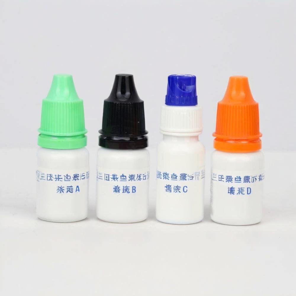
Apply Solution A to cover the entire blood film and fix for 2-3 minutes.
Add an equal amount of Solution B, gently shake the slide to mix Solution A and Solution B thoroughly. A bright coppery film will appear (staining process).
Let it stand for staining for 3-5 minutes.
Rinse slowly from one end of the slide with distilled water (note: do not pour off the stain first or rinse directly on the blood film). Allow to air dry before microscopic examination.
Interpretation of Test Results:
White blood cells: Yellow – red.
Polymorphonuclear granulocytes: Nucleus is dark purple; granules are red – light purple; cytoplasm is cyan – pink.
Eosinophils: Nucleus is blue; granules are red to orange – red; cytoplasm is blue.
Basophils: Nucleus is purple to dark blue; granules are dark purple.
Lymphocytes: Nucleus is dark purple; cytoplasm is sky blue.
Platelets: Granules are purple to dark purple.